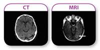
Diagnostic
imaging is a technique of taking images of the different internal parts
of the body for medical purposes. It can be done for diagnosing or
examining a certain disease. MRI and CT-Scan are the two common types of
medical imaging to study the brain. While both have similar functions,
it is important to know what distinguishes one from the other.
MRI or CT-Scan
While
both types of diagnostic imaging are used to examine the brain, the use
of either one depends on the purpose. Each procedure has its advantages
and disadvantages. One may be better to get a quick scan while the
other is better in examining a specific part of the head. Also, the age
of the patient is a factor in determining which medical imaging
technique to perform. In any case, the doctor will determine which test
the patient will need. Sometimes, the doctor will order both.
Why the CT-Scan?
The
CT-Scan or CAT Scan stands for Computed Tomography. It is a diagnostic
imaging process that gets images faster, which makes it the go to choice
for trauma and neurological emergencies. It is considerably cheaper.
CT-Scan is also less sensitive to patient movement as it can be
performed quickly; this is advantageous for patients that are
claustrophobic.
The
CT-Scan evaluates the cortical bone, detects calcification and can
detect metal foreign objects in the body. There’s no risk involved even
if the patient has an implanted medical device like nerve stimulators
and pacemakers.
CT
Scans are best used with small bone structures, brain trauma, pelvis,
chest, spinal column and the abdomen. Sometimes, the patient is injected
with barium sulfate to assist in making certain parts of the body
appear clearer in the image scan.
Why the MRI?
MRI
or Magnetic Resonance Imaging is a diagnostic imaging process that
doesn’t use ionizing radiation. It is highly preferred to be used on
children and patients that need to undergo a number of imaging exams.
MRI is better for evaluating soft tissue contrast and determining brain
abnormalities. Further, it evaluates structures that are not so clear in
a CAT Scan due to the bone artifacts. This can be done without
physically moving the patient.
The
MRI is best used to assess torn rotator cuffs, torn knee cartilage,
torn ligaments, hip problems and herniated disks. This procedure can
take 30 to 90 minutes.
Only a doctor
can determine which medical imaging process will best suit the condition
of the patient. Hospitals and clinics will always require a written
order from the doctor before approving the procedure to be performed.
Vista Clinical Diagnostics
2727 Martin Luther King Jr. Blvd #220, Tampa, FL 33607
813-964-6623
http://www.vista-clinical.com/


No comments:
Post a Comment